Amoebic liver abscess in an adult Nigerian: A case report
Obiozor AA1, Obiozor AC2
Abstract
Background: Amoebic liver abscess (ALA) is a parasitic infection caused by Entamoeba histolytica. ALA is a significant health concern, particularly in regions with poor sanitation and hygiene practices. Radiological imaging plays a crucial role in the diagnosis and management of ALA.
Methods: We present a case report of a 40-year-old male patient with ALA. The patient presented to a radio diagnostic centre in Umuahia with right upper quadrant abdominal pain, fever, diarrhea and weight loss. Radiological evaluation was performed using abdominal ultrasound and contrast-enhanced CT scan. The imaging findings were correlated with clinical presentation and laboratory investigations.
Results: The radiological findings revealed a focal hypoechoic lesion in the right hepatic lobe with ill-defined margins and internal echoes suggestive of liquefaction. The contrast-enhanced CT scan confirmed the presence of a well-defined, hypodense lesion with peripheral enhancement and central hypoattenuation. No evidence of biliary ductal dilatation or vascular invasion was observed. Based on the findings, a diagnosis of ALA was made, and the patient was initiated on intravenous metronidazole therapy. Follow-up imaging demonstrated a decrease in abscess size and resolution of internal echoes, indicating a positive response to treatment.
Conclusion: Radiological imaging, including ultrasound and CT scan, plays a pivotal role in the diagnosis and management of ALA. The characteristic imaging findings, such as hypoechoic lesions with internal echoes and peripheral enhancement with central hypoattenuation, aid in differentiating ALA from other hepatic lesions. Early diagnosis and prompt initiation of appropriate therapy are crucial for favorable patient outcomes. Radiological follow-up allows monitoring of treatment response and guides further management decisions.
Keywords: Ultrasound; Contrast Enhanced CT scan, Intravenous Metronidazole, Umuahia.
Introduction
Amoebic liver abscess (ALA) is a significant infectious disease caused by the protozoan parasite Entamoeba histolytica. Two distinct species of Entamoeba are now recognized: Entamoeba histolytica and Entamoeba dispar. E. histolytica is the cause of dysentery, colitis, and ALA, whereas E. dispar has clinically never been associated with disease.1 It is a common tropical and subtropical infection, particularly prevalent in regions with inadequate sanitation and poor hygiene practices. ALA is characterized by the formation of abscesses within the liver parenchyma, leading to a range of clinical manifestations.
The infection occurs through the ingestion of contaminated food or water containing the cysts of E. histolytica. Once ingested, the cysts release trophozoites in the large intestine, which can invade the colonic mucosa and spread via the portal circulation to the liver. The trophozoites can cause tissue destruction and trigger an inflammatory response, resulting in the formation of abscesses2.
Clinical presentation of ALA is variable but typically includes symptoms such as right upper quadrant abdominal pain, fever, hepatomegaly, and sometimes weight loss3. The severity of symptoms can range from mild discomfort to severe systemic illness. In some cases, ALA can be asymptomatic, leading to delayed diagnosis and potential complications.
Imaging plays a crucial role in the diagnosis of ALA. Ultrasound is often the initial imaging modality of choice due to its availability, cost-effectiveness, and ability to detect characteristic findings such as hypoechoic lesions with internal echoes suggestive of liquefaction4. Computed tomography (CT) scan provides additional details on the size, location, and extent of the abscesses, aiding in treatment planning and monitoring.
Prompt diagnosis and appropriate management are essential to prevent complications such as rupture, peritonitis, or dissemination to other organs. Medical therapy with metronidazole, a nitroimidazole antimicrobial agent, is the standard treatment for ALA. In some cases, large or complicated abscesses may require percutaneous drainage or surgical intervention.
In this case presentation, we discuss the clinical history, radiological findings, diagnosis, and management of a 40yr old patient who presented to radio-diagnostic centre in Umuahia with ALA. The aim is to highlight the importance of timely recognition, appropriate imaging evaluation, and multidisciplinary management approaches to optimize patient outcomes and prevent complications associated with this parasitic infection.
Case report
Patient Information: Age: 40 years old; Gender: Male; Chief Complaint: Right upper quadrant abdominal pain, fever, diarrhoea and weight loss.
Clinical History: The patient presented with a two-week history of right upper quadrant abdominal pain, intermittent fever, diarrhoea and unintentional weight loss. No history of recent travel or exposure to contaminated water was reported. Physical examination revealed tenderness over the right upper quadrant.
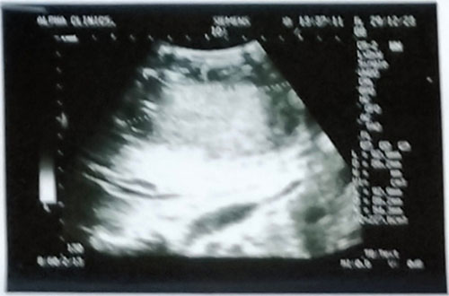
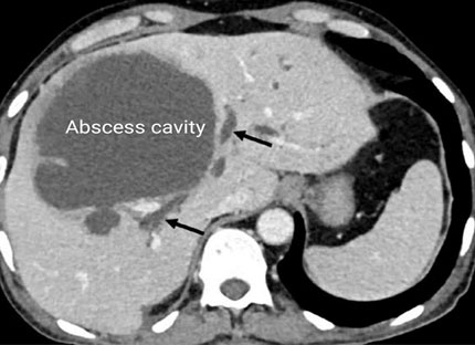
Radiological Findings: An abdominal ultrasound was performed, revealing the following findings: The liver appeared enlarged with a focal hypoechoic lesion measuring approximately 6 cm in diameter, located in the right lobe; the lesion demonstrated an irregular shape with ill-defined margins; internal echoes within the lesion were noted, suggestive of liquefaction; surrounding liver parenchyma showed mild heterogeneous echogenicity. Based on the ultrasound findings, a contrast-enhanced CT scan of the abdomen was performed, which revealed: A well-defined, hypodense lesion with peripheral enhancement in the right hepatic lobe with HU of 20 which is in keeping with an abscess. The lesion displayed central hypoattenuation, indicative of liquefaction with HU of 0. No evidence of intrahepatic biliary ductal dilatation or vascular invasion was observed and no other significant abnormalities were noted in the abdomen or pelvis.
Diagnosis and management: Blood tests on arrival showed leucocytosis (white cell count 31 × 109/L) with markedly elevated inflammatory markers (C-reactive protein 357 mg/L), and deranged liver function (alkaline phosphatase 231 U/L, alanine aminotransferase 64 U/L, bilirubin 56 μmol/L and albumin 26.5 g/L). Blood cultures were negative. No abnormalities were found on colonoscopy and stool ova, cysts and parasite microscopy were negative. Ultrasound guided drainage of the abscess was negative on microscopy and culture, but positive for Entamoeba histolytica.
Based on the radiological findings, laboratory investigations and clinical presentation, a diagnosis of amoebic liver abscess (ALA) was made. The patient was started on metronidazole therapy for amoebic infection. Serial imaging studies were performed to monitor the response to treatment.
Follow-up: Follow-up ultrasound examinations were conducted after one week at intervals to assess the progression of the amoebic liver abscess and response to therapy. The patient's symptoms gradually improved, and subsequent imaging demonstrated a decrease in the size of the abscess cavity and resolution of the internal echoes after one week.

Figure 1: Ultrasound image showing the amoebic liver abscess

Figure 2: Axial CT scan image of amoebic liver abscess
Discussion
Amoebic liver abscess is a common parasitic infection caused by the protozoan Entamoeba histolytica. Imaging plays a crucial role in the diagnosis and management of ALA. Ultrasound is often the initial modality of choice and can detect characteristic findings such as a hypoechoic lesion with internal echoes suggestive of liquefaction. CT scan provides additional details on the morphology, size, and extent of the abscess.
About 10% of the world’s population are infected with E. histolytica, making amebiasis about the third most common cause of death from parasitic diseases. The areas of highest incidence are mostly in developing tropical countries. The Highest endemicity in communities with low socioeconomic status, inadequate sanitation, and overcrowding. Most individuals harboring the parasite are healthy carries who eliminate up to 1.5 x 10⁹ cysts in their daily stools.5
Entamoeba histolytica may be viewed as a cytotoxic effector cell, with an extraordinary capacity of killing various target cells and also a primitive actively phagocytozing eukariotic cell that uses bacteria as a major nutrient source. Initial steps in tissue invasion may be aided by the release of proteases from trophozoites, which are capable of degrading the extracellular matrix components such as fibronectin and laminin and activation of the host complement system. When contact is made, ameba can lyse the target cell, using pore-forming molecules called ameba pores, and possibly phospholipases.6,7
ALA can be found in all age groups but is more frequent in males between the age of 20 and 40 years. Gender differences may be related to alcohol consumption.8 The signs and symptoms of the disease can vary according to the severity of the illness, there are some typical characteristics of this condition: the onset is abrupt with fever between 37 to 40°C, accompanying chills and profuse sweating specially in the afternoon and at night. Almost all patients complain of intense and constant pain in the right upper abdominal quadrant, radiating to the scapular region and right shoulder. When the ALA appears in the left lobe, pain can be felt in the epigastrium and may radiate to the left shoulder.
Abscess sizes may vary considerably, from pinpoint lesions to extremely large masses. During autopsy, the average sizes of the abscesses may range from 5 to 15 cm. They are localized preferentially in the right lobe (35%) while multiple abscesses may be about (16%). These variants may have a worse outcome than the classical solitary right lobe abscess.9
It is vital to know that all cases of hepatic amoebiasis most presumably have begun with an intestinal infection, trophozoites or cysts can be demonstrated in the stools very rarely. More than 90% of patients have leukocytosis. Alkaline phsophatase is elevated in nearly half of the patients and is one the most reliable biochemical indicators of ALA. Anti-amebic serum antibodies are present in more than 90%. Serology may be negative during the first week after onset; titers reach a peak by the second or third month, decreasing to lower but still detectable levels by nine months. Although a variety of test is available, indirect hemagglutination (IHA) and enzyme-linked immunoabsorbent assay (ELISA) are the most used. IHA is the most sensitive and specific method, with 85 to 95% of patients testing positive. A cut-off value of 1:512 is considered diagnostic. A commercially available test, which detects circulating E. histolytica Gal/GalNAc lectin antigen is now available, which is positive in almost all patients which ALA tested prior to treatment, and becomes negative two weeks after the beginning of anti-amebic treatment.10
Ultrasonography is the first imaging modality of choice in the diagnosis of ALA. It is cost-effective, readily available and non-ionizing as compared with CT scan. A space-occupying lesion is seen in 75 to 95% of patients. In ultrasonography, the space -occupying lesion is round or oval in shape with well-defined margins with lack of prominent peripheral echoes. The lesions are primarily hypoechoic.9
The patient in this case responded very well to metronidazole which is the drug of choice in the treatment of uncomplicated ALA. The recommended oral dosage is 1 gr orally b.i.d for 10 to 15 days in adults and 30 to 50 mg/kg/d for 10 days in three divided doses for children, when it is used intravenously, 500 mg every 6 hours for adults and 7.5 mg/kg every 6 hours for children during 10 days. Other nitroimidazoles are also effective as tissue amebecide; these include mainly tinidazole, ornidazole, at a dose of 2 gr orally daily for 10 days.10
Ultrasound guided percutaneous aspiration used mostly as a diagnosis tool may occasionally help to accelerate recovery associated with medical treatment. This procedure should only be attempted if the clinician suspects that there is a great risk of rupture or when response to drugs has been slow or when an association with pyogenic infection is suspected.11 The surgical drainage of an uncomplicated ALA is rare, if ever, indicated. Surgical intervention may be necessary for complications of liver abscesses, including rupture into intra-abdominal and/or thoracic surrounding organs.
There is ongoing studies on the development of a vaccine to prevent amebiasis in high risk populations. The E. histolytica Gal/GalNAc is a particularly attractive vaccine candidate for several reasons: it is an antigenically conserved surface molecule is distinct isolates of E. histolytica, it is the major antigen recognized by humoral system, it plays a key role in adherence of the parasite to host cells, and stimulates proliferation of amebicidal immune peripheral lymphocytes and production of protective cytokines.12
Work is also on going on the use of oral vaccination with attenuated Salmonella typhymurium expressing a serine rich E. histolytica protein (SREHP). This represents a potential candidate vaccine against amoebiasis which is suitable for testing in humans.13
Summary: Management of ALA typically involves a combination of medical therapy and ultrasound guided percutaneous drainage for large or complicated abscesses. Metronidazole is the drug of choice for amoebic infections. Serial imaging helps evaluate the response to treatment and guide further management decisions.
Conclusion
This case report describes the imaging findings, diagnosis, and management of a patient with amoebic liver abscess. Radiological studies, such as ultrasound and CT scan, are valuable tools in the evaluation and follow-up of ALA. Early diagnosis and appropriate treatment are crucial in achieving favorable patient outcomes.
References
- Ackers J, Clark CG, Diamond LS. Entamoeba taxonomy. Bull World Health Org 1997; 72: 97-100
- Akgün Y, Taçyιlιdιz ÏH, Çelik Y. Amoebic liver abscess: changing trends over 20 years. World J Surg 1999;23:102-6.
- Donovon AJ, Yellin AE, Ralls PW. Hepatic abscess. World J Surg 1991;15:162-9.
- Petri WA (Jr), Singh U. Diagnosis and management of amebiasis. Clin Infec Dis, 29 (1999), 117-1125
- Beaver PC, Jung RC, Sherman HJ. Experimental Entamoeba histolytica infections in man. Am J Trop Med Hyg 1956; 5: 1000-1009
- Leippe M, Amoebapores. Parasitol Today 1997;13:178-183
- Olivera MA, Torre A, Kershenobich D, Liver abscesses. Disease of the liver: Schiff’s., 9th, Lippincott Williams&Wilkins, (2002)
- Seeto RK, Rockey DC. Amoebic liver abscess: Epidemiology, clinical features, and outcome. West J Med 1999; 170:104-109
- Muñoz LE, Botello MA, Carrillo O. Early detection of complications in amebic liver abscess. Arch Med Res 1999; 23:251-253
- Rosenblatt JE, Edson RS. Metronidazole. Mayo Clin Proc 1987; 62: 1013-1017
- Rajak CL, Gupta S, Jain S. Percutaneous treatment of liver abscesses: Needle aspiration versus catheter drainage. AJR. Am J Roentgenol 1998; 170: 1035-1039.
- Gaucher D, Chadee K. Immunogenicity of and optimized Entamoeba histolytica Gal-Lectin DNA vaccine. Arch Med Res 2000; 31: S307-S308.
- Zhang T, Stanley SJ (Jr). Progress in an oral vaccine for amebiasis. Expression of the serine rich Entamoeba histolytica protein (SREHP) in the avirulent vaccine strain Salmonella typhyx 4297/cya, crp, asd): safety and immunogenicity in mice. Arch Med Res 1997; 28: S269-S271.