Diabetes and Cervical Necrotizing Fasciitis: A Case Report
Uwanuruochi VN1, Ogba JA2, Iwegbu IO3, Uwanuruochi K4, Udeke U5
Introduction
Cervical necrotizing fasciitis in diabetics is under-reported in the literature. We present a case report and discuss salient points in its management.
Case report
A 52 year old man, ME presented at the dental clinic with toothache and swelling in the lower cheek, neck and chest region of one week duration. He is diabetic for 2 years on treatment with Metformin 500mg BD, but not regular with medications and follow up.
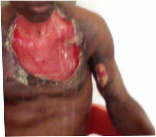
On examination he had a diffuse swelling in the left lower cheek extending to bilateral submandibular regions, (fluctuant and firm in some regions) and the neck as well as upper chest wall. The skin over the neck and chest wall appeared reddish (Figure 1).

He had trismus and an impression of Ludwig’s angina in poorly controlled Type 2 diabetes mellitus with neck cellulitis was made.
Extraction of the lower molar, incision and drainage, and debridement of the left submandibular site was performed on the day of presentation, after which he was placed on parenteral analgesics (intramuscular piroxicam 20mg stat and intramuscular diclofenac 75mg 12 hourly for 5 days), Vitamin C 200mg 8 hourly for 2 weeks and 0.2% Chlorhexidine mouth wash 12 hourly for 2 weeks. In addition, Metformin was replaced with subcutaneous Humulin 70/30 at an initial dose of 10 IU am and 6 IU pm (20-30mins before meals) and empirical antibiotics were given (IV Rocephin 2g stat, then 1g 12 hourly and IV Metronidazole 500mg 8 hourly for 5 days respectively).
Laboratory investigations showed reduced hemoglobin (11.2g/dL), elevated total white blood cells (13.3 × 10⁹ u/L (4-12)) with relative monocytosis (1.3 × 10⁹ u/L (0.1-1)) and neutrophilia (10.4 × 10⁹ u/L (2-8)). Serum electrolytes and creatinine were within normal range, but urea was mildly raised, 54mg/dL(13-43). Urinalysis showed glycosuria. Fasting lipid profile was normal except for Triglyceride which was markedly elevated 235mg/dL (0 -150). His liver function test showed derangements in alkaline phosphatase 233.9 u/L (40-120), reduced albumin 19.6g/dL (35-50), increased globulin 44.8g/dL(0.9-2.7and raised total bilirubin 14umol/L (2-4). Echocardiography revealed mild left ventricular dysfunction.
Incision and drainage was performed several times for pockets of abscesses which had tracked under the skin, with regular and aggressive debridement, hence expanding the ulcer. Patient was initially started on broadspectrum antibibiotics (Ceftriaxone and Metronidazole and later changed based on results of serial wound swab microscopy, culture and sensitivity. Anterior chest wall wound was dressed twice daily with honey and Betadine-soaked gauze (Figure 2).

Three months later patient was stable and was worked up for split thickness skin graft of the anterior chest wall on account of the wide necrosis of the skin that had occurred over it.
Discussion
Necrotizing fasciitis (NF) is a potentially fatal soft tissue infection characterized by excessive necrosis and gas formation in the subcutaneous tissue and fascia.1 It predominantly affects the tissues of the abdominal wall, the perineum and the extremities, but can also be seen in the maxillofacial region.2 NF of the neck is rare and usually occurs secondary to dental infection, gingivitis, or pulpitis. However, any infection that can cause deep neck infection can cause necrotizing fasciitis. The infection starts in the superficial fascia. Enzymes and proteins released by the responsible micro-organisms cause necrosis of fascial layers. Horizontal spread of infection may not be clinically apparent on the skin surface and hence diagnosis may be delayed. This infection spreads along the fascial planes with rapid progression and systemic toxicity, due to the presence of polymicrobes both aerobes and obligate anaerobes, and the synergistic effect of the bacterial enzymes.3-5
In Namibia, Africa, only 1 case has been reported.6 NF occur in 0.4 per 100,000 people annually in the United States.7 Less than 40 cases of cervical NF have been reported worldwide.8 In Nigeria, sixteen cases were reported between 1990-2000 at OAU Ife.5
This is the first report of this condition in Federal Medical Centre Umuahia, South East Nigeria
The most common organisms in polymicrobial NF are combinations of Staphylococci (esp. Staph. Epidermidis) Enterococci, Enterobacteriaceae species, anaerobic gram-positive cocci, Clostridium species and Beta-haemolytic streptococci namely Group A streptococci or Streptococcus pyogens.
Odontogenic infections or procedures are the most important risk factor indicated in Cervical NF in Diabetes Mellitus. In diabetes mellitus, the immune system is suppressed due to the lazy leukocyte syndrome. The pathogenesis is not clear, but there is complete loss of local and immune response due to aberrant chemotaxis, increased leukotriene production, lysosomal enzyme release and production of reactive oxygen species. This increases susceptibility to infections. Following odontogenic infections or oral procedures, there is a loss of protective barriers and bacteria enters the body and infect the muscle fasciae. The first organism that gets into the tissues are aerobes, most notably Streptococcus spp and Staphylococcus aureus. They bind to host cells with M-proteins and Hyaluronic acid capsule, escaping phagocytosis. These organisms then invades tissues reducing oxygen concentration by redox potential and favouring growth and proliferation of anaerobic organisms. The anaerobes produce short chain fatty acids such as butyric acid and propionic acid which inhibit phagocytosis of aerobes. The aerobes grow, proliferate and destroy more tissues; (microbial synergism). The organisms then produce exotoxins such as streptolysin which cause blood clot formation or stimulates programmed cell death.
There is also activation of complement pathway, the bradykinin-kallikrein system, and the coagulation cascade, worsening small vessel thrombosis and tissue ischemia. The blood clot formed blocks tiny blood vessels, causing tissue ischemia in the epidermis, dermis, subcutaneous fat, muscle fasciae and muscles. Ischemia leads also to destruction of nerve endings, loss of sensation and further destruction of tissues, decrease in blood flow to tissues and more tissue death. They also release massive amount of hemolysin, DNAse, protease, and collagenase that destroy the skin and surrounding tissues with progressive coagulation necrosis. Also, the inflamed cells release more cytokines that stimulate more inflammatory cells. The cytokines leads to most of the clinical features seen such as SIRS resulting in shock, organ failure, myocardial depression and death
The frequency of NF has been on the rise because of an increase in immunocompromised patients with diabetes mellitus (DM), cancer, alcoholism, vascular insufficiencies, organ transplants, human immunodeficiency virus infection, or neutropenia. DM is a common underlying disease in NF patients, accounting for 44.5–72.3 % in various series.6 Diabetic patients exhibit impaired cutaneous wound healing and increased susceptibility to infection, which may affect the course of soft-tissue infections.7
The mortality rate is 15%–40%. Successful treatment of this life-threatening condition depends on early diagnosis, aggressive surgical intervention and supportive therapy such as appropriate antibiotics, airway maintenance and adjunctive hyperbaric oxygen therapy.8
This aggressive approach contributed to a good outcome in our case, however we did not use hyperbaric oxygen to treat the patient as he improved on the treatment given; also we do not have facility to treat patient with hyperbaric oxygen.
References
- Lin C, Yeh FL, Lin JT, et al. Necrotizing fasciitis of the head and neck: an analysis of 47 cases. Plast Reconstr Surg. 2001; 107(7): 1684–1693. doi: 10.1097/00006534-200106000-00008
- De Backer T, Bossuyt M, Schoenaers J. Management of necrotizing fasciitis in the neck. J Craniomaxillofac Surg. 1997;24:366–71.
- Morgan MS. Diagnosis and management of necrotizing fasciitis: A multiparametric approach. J Hosp Infect. 2010;75:249–257. doi: 10.1016/j.jhin.2010.01.028.
- Levine EG, Manders SM. Life-threatening necrotizing fasciitis. Clin in Dermat. 2005;23:144–147. doi: 10.1016/j.clindermatol.2004.06.014.
- Ndukwe, K. C., Fatusi, O. A., & Ugboko, V. I. (2002). Craniocervical necrotizing fasciitis in Ile-Ife, Nigeria. The British journal of oral & maxillofacial surgery, 40(1), 64–67. https://doi.org/10.1054/bjom.2001.0715
- Goh T, Goh LG, Ang CH, Wong CH. Early diagnosis of necrotizing fasciitis. Br. J. Surg., 101 (1) (2014), pp. e119-e125, 10.1002/bjs.9371
- Giaccari A, Sorice G, Muscogiuri G. Glucose toxicity: the leading actor in the pathogenesis and clinical history of type 2 diabetes - mechanisms and potentials for treatment. Nutr. Metab. Cardiovasc. Dis., 19 (5) (2009), pp. 365-377, 10.1016/j.numecd.2009.03.018
- Poromanski I. Developing a tool to diagnose cases of necrotizing fasciitis. J Wound Care. 2004;13(8):307–310.